Working Hours - Mon - Sat 9.00 am to 7.30 pm - 0495 - 2740321, +91 9747580707
Showing 41–51 of 51 results
This succinct yet complete presentation of radiologic diseases includes all relevant findings and differential diagnoses for every body region. Similar to previous versions, the fourth edition is organized by body system and is presented in an outline format with line drawings and mnemonics for remembering diagnoses.
Offering lower toxicity and higher accuracy than conventional therapies, this source offers illustrative coverage of this new method to treat tumors associated with brain, breast, lung, and neuroendocrine cancers. Accompanied by a CD offering color images, radiolabeling procedures, and tips on radiopharmceutical administration, this source will offer comprehensive and expert guidance on this burgeoning therapeutic technique.
Breast cancer is the leading cause of cancer-related deaths in women, and its prevalence has been steadily rising in recent decades. This book describes morphologic and kinetic signs that are important in the analysis of breast MR images before and after contrast administration and in various pulse sequences. It will help broaden the clinical application of MRM so that as many physicians as possible can make more accurate diagnoses.
The first Amirsys publication devoted solely to genitourinary cancers has finally arrived. In Specialty Imaging: Genitourinary Oncology, Dr. Akram Shaaban and team skillfully guide clinicians through the imaging appearances, staging, and treatment techniques for cancers of the kidney, bladder, adrenal glands, urethra, testicle, and prostate. The 130 pages of fast-reading, bulleted text are enriched with well over 450 vivid images, each of which identifies and explains clinically significant factors. The recently published Seventh Edition AJCC cancer staging tables are included to aid the staging discussions, providing definitions for TNM and AJCC prognostic groups for all genitourinary cancers. This is a must-have reference for radiologists, oncologists, and others whose work quality depends on an accurate understanding of genitourinary system cancers.
Written by recognized experts, this volume is a comprehensive reference on the use of advanced imaging techniques in the diagnosis and management of spinal trauma. In one cohesive source, the book brings together information on state-of-the-art clinical imaging—including multidetector CT and high-field MRI techniques—and the pathophysiology, neurologic evaluation, medical management, surgical treatment, and postoperative assessment of spine trauma and spinal cord injury. Also included are cutting-edge reviews of experimental imaging techniques and their applications and experimental therapies such as neurotransplantation. More than 700 illustrations—including 180 in full color—complement the text.
All the guidance you need to enhance your understanding and clinical application of ultrasound
Includes DVD with video of key techniques
Surgical and Interventional Ultrasound offers a thorough survey of image-guided treatments in the OR, in the endoscopy suite, and at the bedside. This one-stop clinical companion spans virtually every kind of surgical and interventional specialty that utilizes ultrasound and delivers high-yield perspectives on using these techniques to ensure accurate clinical decision making.
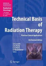
This book is unique in detailing in depth the technological basis of radiation therapy. Compared with the previous edition, all chapters have been rewritten and updated. In addition, new chapters have been included on various topics, including the use of imaging in treatment planning, second malignant neoplasms due to irradiation, and quality assurance in radiation oncology. The book is divided into two sections. The first covers basic concepts in treatment planning and explains the various approaches to radiation therapy. The second part documents the practical clinical applications of these concepts in the treatment of different cancers.
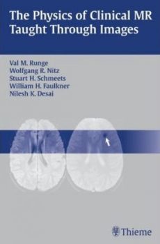
Portions of the book have been presented at both the RSNA (Radiological Society of North America, 2003) meeting and the ARRS (American Roentgen Ray Society, 2004) meeting. Awards were given to one of the two exhibits shown at each meeting, with the award at the ARRS being very prestigious (a ‘silver medal’). At the 2004 ARRS meeting there were 229 exhibits presented. One gold medal, three silver medals, six bronze medals and 30 certificates of merit were awarded. The exhibits were also chosen for submission to Radiographics (which was declined, as there were copyright issues). Other sections of the book have been submitted for presentation at the 2004 RSNA meeting, and if accepted there, will also be submitted to the 2005 ARRS meeting.
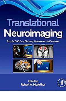
ranslational Neuroimaging: Tools for CNS Drug Discovery, Development and Treatment combines the experience of academic, clinical and industrial neuroimagers in a unique collaborative approach to provide an integrated perspective of the use of small animal and human brain imaging in developing and validating translational models and biomarkers for the study and treatment of neuropsychiatric disorders. Translational Neuroimaging: Tools for CNS Drug Discovery, Development and Treatment examines the translational role of neuroimaging in model development from preclinical animal models, to human experimental medicine, and finally to clinical studies. The focus of this book is to identify and provide common endpoints between species that can serve to inform both the clinic and the bench with the information needed to accelerate clinically-effective CNS drug discovery. This book covers methodical issues in human and animal neuroimaging translational research as well as detailed applied examples of the use of neuroimaging in neuropsychiatric disorders and the development of drugs for their treatment. Translational Neuroimaging: Tools for CNS Drug Discovery,
Ultrasonic energy can be applied to surgical instruments for cutting, dissecting, and coagulating vessels and/or tissue. This book presents the use of ultrasound energy as it is used in UltraCision products, for use in surgery on many parts of the body.
Principles of UltraCision technology: The instruments are attached to a hand piece, which is connected to a generator. The ultrasonic system is situated inside the hand piece and transforms the electric energy into mechanical movement, producing in this way longitudinal vibrations of the blades at approx. 55 kHz per second. The movement of the blades causes collagen molecules to vibrate inside the tissues, thus forming a coagulum upon their denaturation. This method is especially useful in parenchymatous organs, where dissection can be performed cleanly and with little or no bleeding.
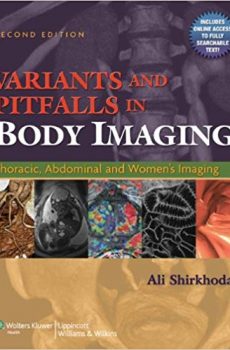
Variants and Pitfalls in Body Imaging, Second Edition is the key to identifying features on images that can impede accurate diagnosis, particularly normal anatomic variants and technical artifacts that mimic pathology. Covering the abdomen, pelvis, and thorax and all current imaging modalities, this sourcebook explains how to differentiate normal anatomic variants, technical artifacts, and other diagnostic pitfalls from pathologic conditions.
Organized by site for easy reference, the book covers CT, MRI, ultrasound, and nuclear medicine. This edition includes advanced technologies such as multidetector CT scanning for cardiovascular imaging, CT and MR enterography for enterocolitis, virtual colonoscopy, CT and MR urography, prostate and breast MR imaging, and PET/CT scanning. Well-respected radiologists walk the reader through specific body areas, describing problems, solutions, and relevant anatomy. Users will come away with a clear understanding that will yield on-target assessments for every patient.
Calicut Books
Opp.Govt Mental Health Centre,
P.O Kuthiravattom,
Calicut - 673016